Information in image scans for medical applications (CT scan, MRI, ultrasound etc.) contain vastly rich information that helps in patient risk management and treatment. With the imaging sciences team at HeartFlow, I developed artificial intelligence (AI) based algorithms that can quantify the degree of vessel narrowing, an important metric to diagnose coronary artery disease. Our work demonstrates a better agreement than state-of-the-art techniques against the read of expert radiologists. [2]
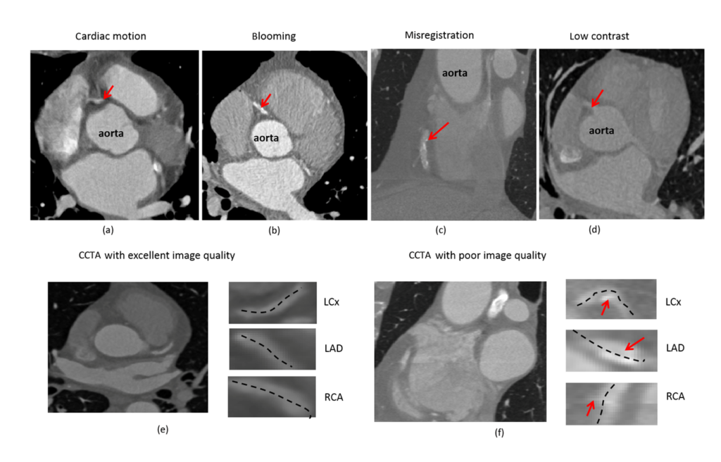
Recently, I developed AI based algorithms for automated assessment of image quality in CT scans. The method synthesizes local image intensity features with a cross-correlation based mis-registration metric and global factors such as contrast-to noise ratio, trained against annotated information from hundreds of scans to quantify image quality. In a recent publication in European Radiology [1], we demonstrated that this algorithm works as good as expert radiologists.
Relevant publications
- Nakanishi, R., Sankaran, S.* et al., Automated Estimation of Image Quality for Coronary Computed Tomographic Angiography using Machine Learning, European Radiology, Vol.28/9, pp. 4018-4026, 2018 (* indicates equal contribution).
- Sankaran, S., et al., HALE: Healthy area of lumen estimation for vessel stenosis quantification, Lecture notes in Computer Science: MICCAI, Vol. 9902, pp. 380-387, 2016.
- Sankaran, S., Grady, L., and Taylor, C., Fast computation of hemodynamic sensitivity to lumen segmentation uncertainty, IEEE Transactions on Medical Imaging, 34 (12), pp. 2562-2571, 2015.